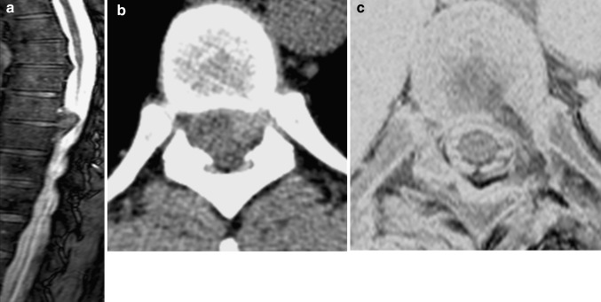
Fig. 5.
Sagittal T2-weighted MRI image showing a thoracic disc herniation at T8–9 with cord compression (a). Axial slice CT scans of T8–9 soft disc herniation (b). A postoperative T2-weighted axial MRI scan showing complete decompression of the T8–9 disc herniation (c) MRI scan obtained on postoperative day 1

