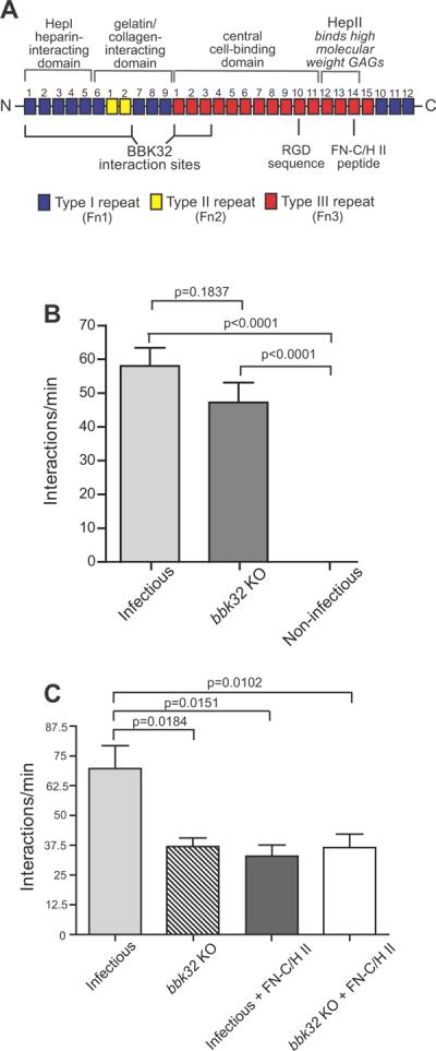
Figure 2. Effect of bbk32 disruption and Fn heparin-binding peptide on microvascular interactions of B. burgdorferi in vivo.

A) Schematic depicting the organization of plasma Fn (not to scale). Major domains are indicated by labels above the schematic. BBK32 interaction sites, and the locations of RGD and FN-C/H II heparin-binding peptides are indicated below the schematic. Numbers above each Fn repeat indicate repeat number, and Fn Type 1, 2 and 3 repeats are color-coded in blue, yellow and red, respectively. Information on Fn structure and the Fn regions where BBK32 interacts are from (Kim et al., 2004, Pankov & Yamada, 2002, Prabhakaran et al., 2009, Raibaud et al., 2005). B) Microvascular interaction rates in skin (transient and dragging interactions combined) are shown for three GFP-expressing B. burgdorferi strains, as analyzed by high acquisition rate spinning disk confocal intravital microscopy. Strains (see also Table S1): 1) GCB966: B31 derivative ML23 (infectious following transformation with a shuttle vector containing bbe22), 2) GCB971: parental with genetic disruption of bbk32 (bbk32 KO), 3) GCB705: non-infectious high passage control (B31-A derivative). A total of 10,387 interactions in 169 venules from 16 mice (n=6 infectious; n=5 bbk32 KO; n=5 non-infectious) were analyzed. Means and standard error bars are indicated for each experimental group. Statistical testing for significant differences among all experimental groups was performed using a non-parametric Kruskal-Wallis ANOVA (overall P value of <0.0001), followed by Dunn's multiple comparison test. *P<0.05, **P<0.01, ***P<0.001. All statistical data for pairwise comparison between groups may be found in Table S4. Microvascular interactions were measured between 5 and 45 minutes after spirochete injection. The average time after injection for recordings made with infectious, bbk32 KO, and non-infectious strains was 22.63−/+1.31, 23.32−/+2.02, and 23.19−/+2.07 min, respectively. C) Microvascular interaction rates in the knee joint are shown for two GFP-expressing B. burgdorferi strains in mice treated or untreated with the fibronectin heparin-binding peptide (FN-C/H II), as analyzed by high acquisition rate spinning disk confocal intravital microscopy. Strains (see also Table S1): 1) GCB966: parental B31 derivative ML23 (infectious following transformation with shuttle vector containing bbe22 and PflaB-gfp), 2) GCB971: GFP-expressing parental with genetic disruption of bbk32. The interactions in 84 venules from 12 mice (n=3 mice/experimental group) were analyzed. Microvascular interactions were measured between 10 and 50 minutes after spirochete injection. Means and standard error bars are indicated for each experimental group. Statistical testing for significant differences among all experimental groups was performed using a non-parametric Kruskal-Wallis ANOVA (overall P value of 0.0114). P-values obtained by two-tailed t-test with correction for unequal variance are indicated for selected pair-wise comparisons. All statistical data may be found in Table S4.
