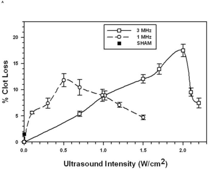
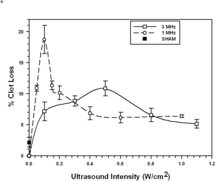
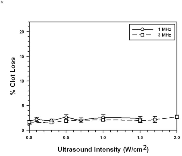
FIGURE 1. Sonothrombolysis efficacy as a function of Ultrasonic Intensity at 1 MHz and 3 MHz (pulsed ultrasound with 100 Hz PRF, 2 ms PD, and 20% duty factor).


Each symbol represents the mean of a minimum of four measurements and the error bars indicate standard deviation. Data points near minima and maxima are the results of six to ten measurements to ensure accuracy.
A. With 1 μm diameter Microbubbles (5.4 × 108 MB/mL).
B: With 3 μm diameter Microbubbles (1.1 × 108 MB/mL)
C: Without Microbubbles
