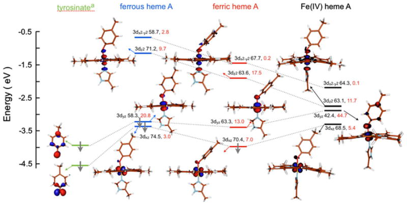
Fig. 12.

DFT-calculated valence β-spin energy-level diagram. Meth-ylphenolate was used as a deprotonated Tyr mimic. Contour plots were generated with Molden. Gray arrows indicate a spin-down electron. The dotted gray lines indicate the contribution of the two deprotonated Tyr ψHOMO in the valence levels of heme A in different oxidation states. The Fe (3d) and deprotonated Tyr contributions to the total Mulliken spin populations are shown next to each level in black and red, respectively. The highest-energy 3dx2−y2 orbital contour has been omitted for clarity. The valence α-spin counterparts of the 3dx2−y2 and 3dz2 orbitals are similar in composition to their β-spin counterparts
