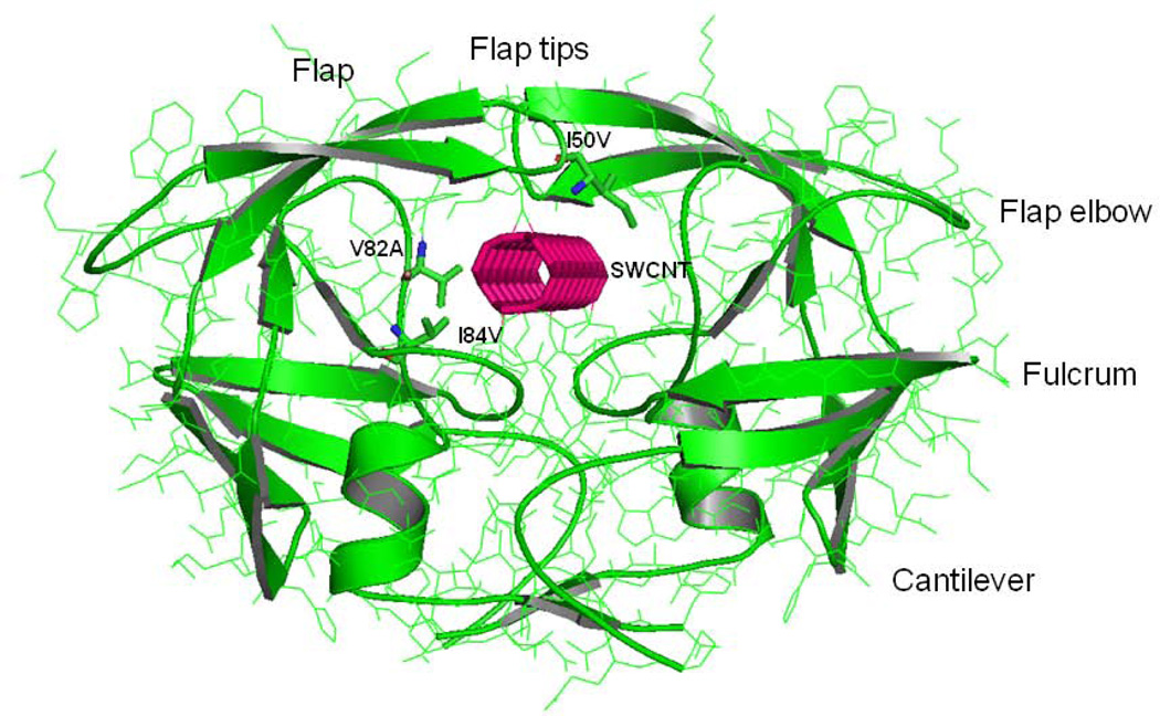
Figure 1.
Configuration of the HIV-PR/SWCNT complex. The HIV-PR is shown in green ribbons for both the chain-A and chain-B. The sites of mutation for Ile50, Val82 and Ile84 are indicated by stick representation. SWCNT is bound in the active site and is labeled, which is represented by solid pink line. Important regions of the HIV-PR like flap, flap elbow, fulcrum and cantilever are also labeled.

