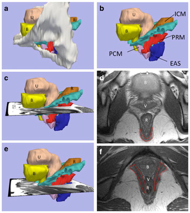
Fig. 1.
Structural orientation to levator ani subdivisions and visibility of the puborectal muscle in MR images at two different levels (chosen from multiple contiguous sections as we followed the puborectalTA muscle from its origin at the pubic bone to its decussation behind the rectum); a lateral view of 3-D model with pubic bones, (B bladder, U uterus, R rectum), b levator subdivisions: without bones (PCM pubococcygeus muscle, ICM iliococcygeus muscle, PRM puborectal muscleTA, EAS external anal sphincter). c Level of the PRM decussation showing the scan plane for d, revealing the MRI at level of the PRM decussation (outlined), e level where the PRM “arms” project towards the pubic bone; f MRI at the level of the PRM “arms” (outlined in red)

