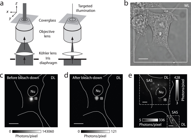
Figure 2. Targeted photo-bleaching of inclusion bodies and selective illumination of sub-cellular regions.
(a) Schematic of the procedure employed for bleaching a sample region containing a strongly fluorescent inclusion body (IB), to reduce its detrimental signal interference with nearby dimmer structures. The IB was first moved to the center of the excitation beam profile. This location was then singled out for photo-bleaching at high intensity (~2.5 kW/cm2 at peak) by narrowing an iris diaphragm in the excitation beam path. (b) White-light (WL) image of a transfected PC12m cell. (c) The diffraction-limited (DL) epi-fluorescence image of eYFP-labeled Htt-ex1 before bleachdown (boxed region in b) was entirely dominated by the IB signal (image recorded without EM gain). Nu = Nucleus. (d) After 12 minutes of bleach-down, the IB's signal had essentially disappeared. (e) Small aggregate species (SAS) were subsequently perceived (shown here with EM gain in a composite image of two regions). The residual fluorescence from the IB still had to be excluded by delimiting the regions of interest with the iris in each case, because the IB brightness slowly recovered over time as eYFP returned from very long-lived dark states. Scale bars: 10 µm (b–d), 5 µm (e).

