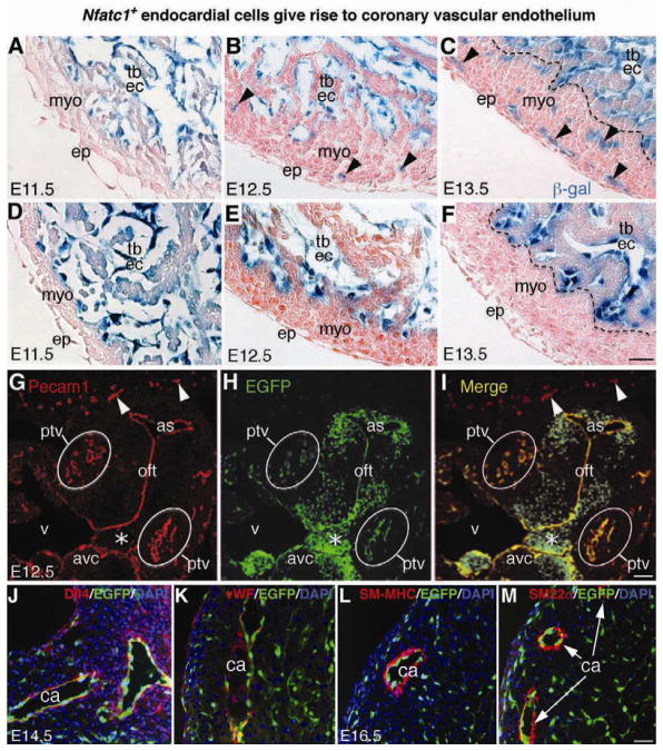
Figure 2. Fate-mapping analysis reveals that Nfatc1+ endocardial cells generate coronary vascular endothelium.
(A–C) X-gal stained E11.5–E13.5 heart sections of Nfatc1Cre;R26fslz embryos show that the β-gal tagged Nfatc1+ endocardial cells reside in the endocardium of the myocardial wall and trabeculae at E11.5 (A), they begin to invade the myocardium at E12.5 (B, arrowheads) and generate networks of coronary plexuses at E13.5 (C, arrowheads). The descendants of Nfatc1+ cells are not present in the epicardium and myocardium (see also Figures S2 and S3).
(D–F) X-gal stained E11.5–E13.5 heart sections of control Nfatc1lacZ-BAC embryos show that the β-gal activities directed by the Nfatc1 promoter/enhancer are restricted to the endocardium and not present in the myocardium and epicardium. Unlike Nfatc1Cre, Nfatc1lacZ does not label the coronary plexuses.
(G–I) Dual fluorescent sections through ventricular (v) outflow tract (oft) of E12.5 Nfatc1Cre;RCEfsEGFP embryos show the EGFP+ endothelial descendants of Nfatc1+ endocardial cells in the Pecam1+ coronary plexuses in the peritruncal region (ptv). The peripheral vessels (arrowheads) expressing Pecam1 but not EGFP are not derived from the Nfatc1+ endocardial cells (see also Figure S4). Mesenchyme of atrioventricular canal (avc, asterisk), derived from the cushion endocardial cells, is EGFP positive but Pecam1 negative.
(J–K) Dual fluorescent sections of E14.5 Nfatc1Cre;RCEfsEGFP heart show EGFP+ endothelial descendants of endocardial cells in the Dll4+ or vWF+ endothelium (red) of the main coronary arteries (ca). The trabecular endocardium is negative for vWF.
(L–M) Dual fluorescent sections of E16.5 Nfatc1Cre;RCEfsEGFP heart show EGFP+ endothelial descendants of endocardial cells in the inner layer of the coronary arteries (ca) surrounded by smooth muscle cells positive for SM-MHC or SM22a (see also Figure S5 and S6). All Bars = 25 μm.

