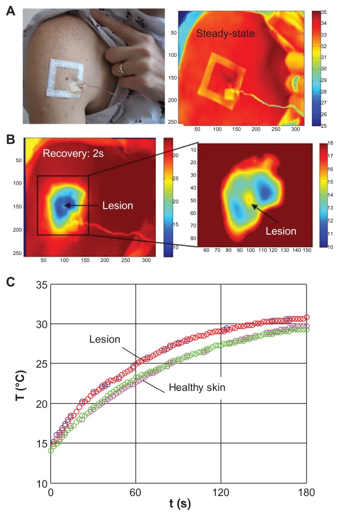
Figure 3.
(A) White light photograph of the larger body surface area with a cluster of pigmented lesions, adhesive window serving as thermal marker and reference IR image of the region at ambient temperature; (B) the same area 2s into the thermal recovery and magnified section of the melanoma lesion and its surroundings; (C) temperature profiles of the lesion and the surrounding normal skin during the thermal recovery process.51–53

