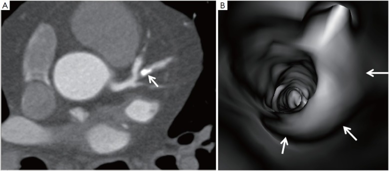
Figure 2.
A calcified plaque is detected at the left anterior descending (arrow in A) with more than 70% lumen stenosis on a 2D axial image. However, virtual endoscopic image confirms that the lumen stenosis is less than 50% (arrows in B). The overestimated stenosis on 2D image is due to blooming artefacts

