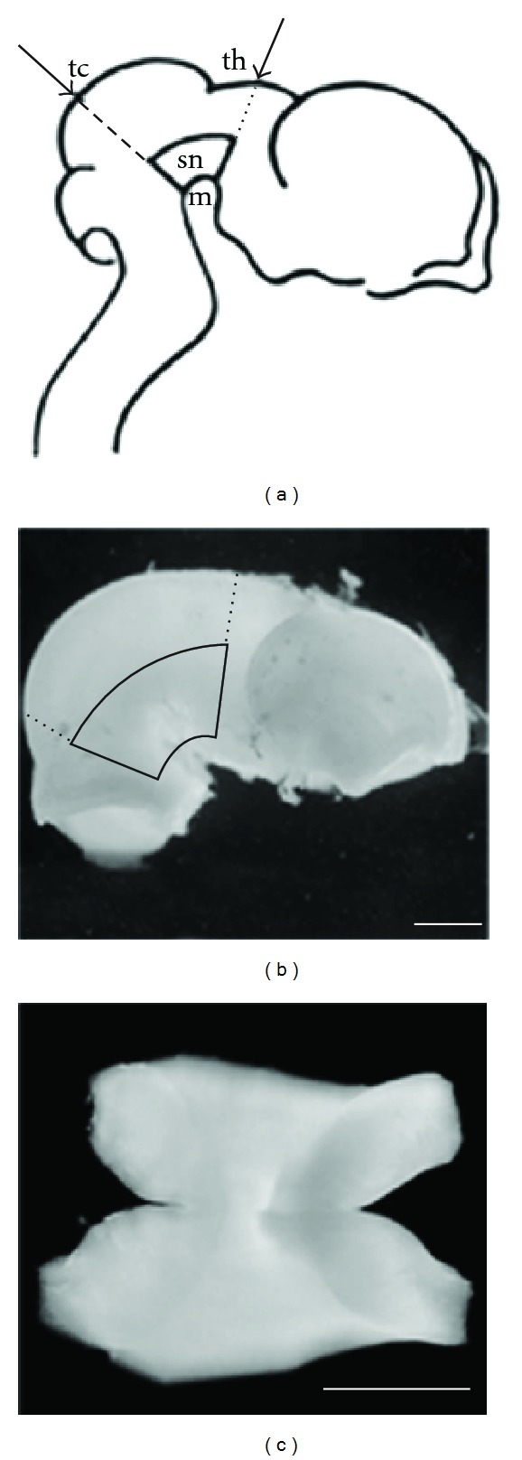
Figure 1.

Dissection of porcine embryonic (E28–30) ventral mesencephalon (VM). (a) Lateral perspective showing the angle of the first two cuts of the dissection, with imaginary lines (arrows) intersecting the dorsal surface of the tectum (tc) and the thalamus (th) (from Dunnett and Bjorklund, 1992). (b) Brain of a 28-day-old embryo with the dissection lines from (a) marked. The area encircled by solid lines represents the VM. (c) The ventral surface of the isolated VM explant. Scale bars: 500 μm.
