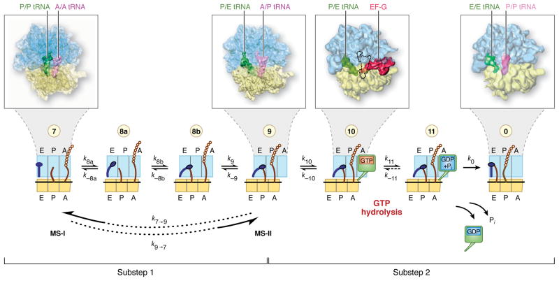
Fig. 1.
The mRNA-tRNA translocation process, and definition of translocation substeps 1 and 2. This diagram presents a sequence of distinct steps during the process of mRNA-tRNA translocation, as characterized by biochemical and structural studies. The blue feature on the left symbolizes the L1 stalk in its “open” and “closed” conformations. The green box on the right in states 10 and 11 stands for EF-G in GTP and GDP forms. Substep 1 requires the rotation of the small subunit body (symbolized by a shift of the yellow box with respect to the blue box) and effects the translocation of the tRNA on the large subunit, through the formation of hybrid A/P and P/E configurations. Substep 2, reached upon binding of EF-G followed by GTP hydrolysis, effects the translocation of mRNA and the tRNAs bound to it with respect to the small subunit. This review focuses on substep 1, which involves several structural intermediates. (Note: In state 10, it has not been possible to trap a complex containing P/E-tRNA, A/P-tRNA and EF-G all in one complex for cryo-EM. For illustration of the general binding position of EF-G on the ribosome, the cryo-EM map depicted here shows a complex that lacks the A-site tRNA and thus allows EF-G to bind in the GTP form. The predicted position of A/P-tRNA in a hypothetical authentic EF-G-bound pretranslocational complex is indicated by an outline. In such a complex, the steric conflict with domain IV of EF-G is presumably resolved by the positional flexibility of this domain known for the free form of EF-G [13]) (Adapted from [2]).

