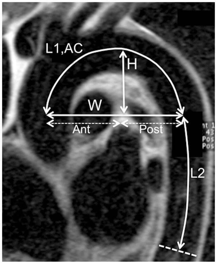
Figure 1. Aortic Arch Geometry assessment with MRI.

Sagittal oblique view of the thoracic aorta using a spin echo black blood MRI acquisition illustrating aortic measurements: L1: length of the aortic arch, AC: average arch curvature, H: arch height, W: arch width, Ant: anterior arch width, Post: posterior arch width. L2: length of the descending aorta.
