
Figure 2.
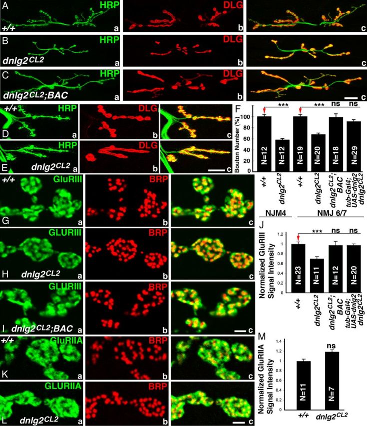
Synaptic bouton growth at NMJs is reduced in dnlg2 mutants. A–E, Confocal images of NMJ6/7 (A–C) and NMJ4 (D, E) from abdominal segment 3 of third-instar larvae labeled with anti-HRP (green) and anti-Dlg (red). Compared with wild-type NMJ6/7 (A), dnlg2 homozygous mutants (B) show reduced NMJ expansion and fewer boutons. This phenotype is rescued by a BAC transgene containing dnlg2 genomic region (C). At NMJ 4, compared with wild type (D), dnlg2 homozygous mutants (E) have fewer boutons, which appeared to be merged. F, Quantifications of type Ib and Is bouton number at NMJ6/7 and type Ib bouton number at NMJ4 adjusted to wild-type bouton number. The bouton number deficits in dnlg2 mutants are rescued by BAC transgene or by ubiquitous Dnlg2 expression using tubP-Gal4. G–I, Confocal images of synaptic boutons at segment 3 NMJ6/7 labeled with postsynaptic marker, GluRIII (green) and AZ marker, Brp (red). The alignment of presynaptic and postsynaptic areas appears to be unaffected in dnlg2 mutants (H, c). However, the levels of GluRIII in dnlg2 mutants (H, a) are significantly reduced. This phenotype is rescued by the BAC transgene (I, a). J, Quantification of GluRIII signal intensity with 3D reconstructed confocal images using Volocity software also reveals reduction in intensity in dnlg2CL2 mutants. K, L, Confocal images of synaptic boutons at NMJ6/7 labeled with postsynaptic marker, GluRIIA (green) and BRP (red). The alignment of GluRIIA with AZ and the levels of GluRIIA are unaffected in dnlg2 mutants (L). M, Quantification of GluRIIA signal intensity shows slight but not significant increase in dnlg2 mutants compared with wild type. Error bar indicates SEM;***p < 0.001; **p < 0.01; *p < 0.05 (Student's t test). Scale bars: A–E, 20 μm; G–L, 2 μm.