
Fig. 1.
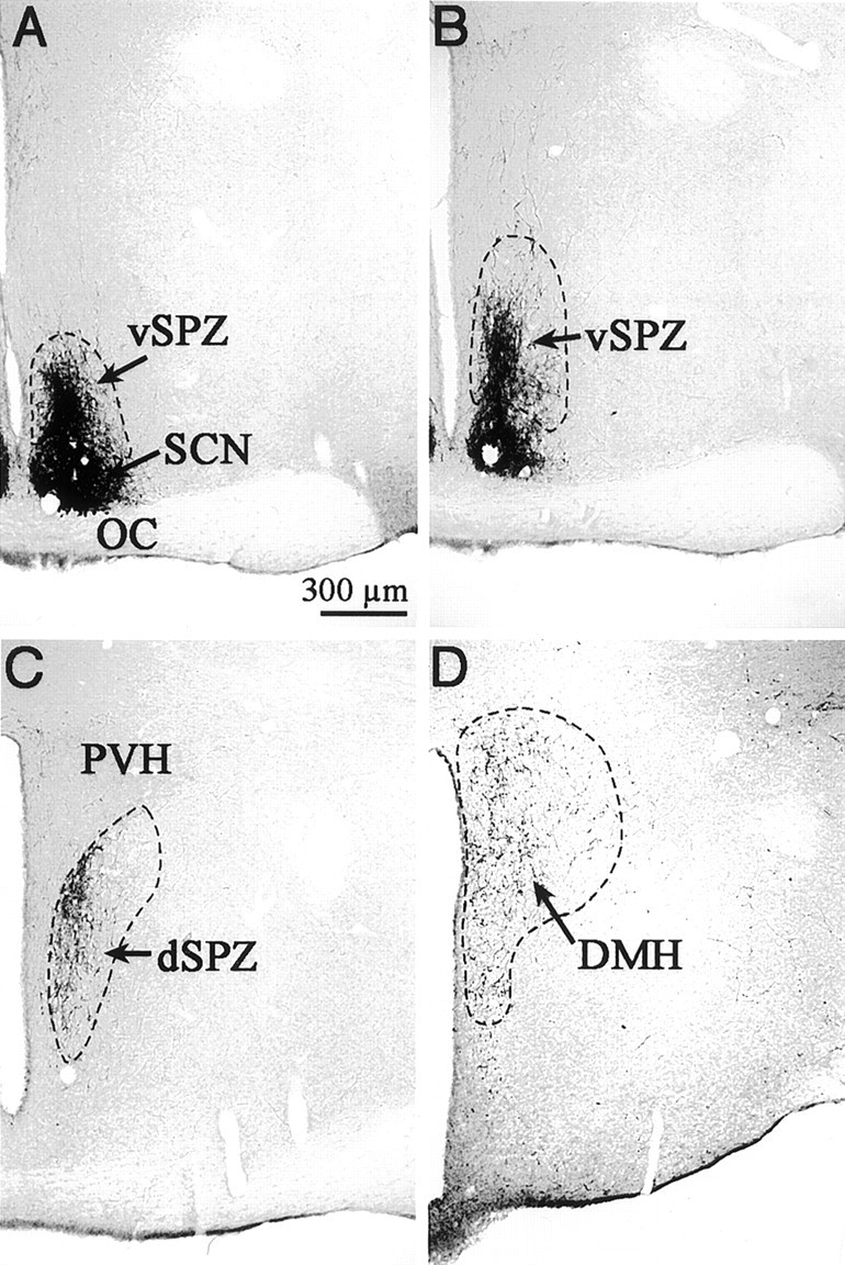
Photomicrographs showing VIP-ir axons and terminals that define the subparaventricular zone (SPZ) from rostral to caudal levels. A, B, The ventral SPZ demonstrated by a column of VIP-ir axons leaving the dorsal margin of the suprachiasmatic nucleus (SCN).C, The dorsal SPZ, ventral to the paraventricular nucleus (PVH). Notice the VIP-ir terminals in the medial PVH. D, The continuation of VIP-ir terminals from the SCN into the rostral DMH.