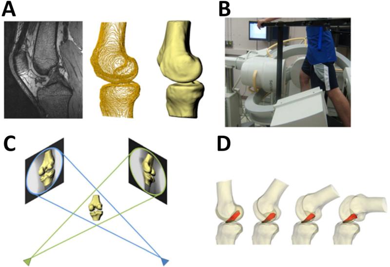
Figure 3.
Multi-planar, high resolution MR imaging was used to create 3D models of the knee, including the attachment sites of the ACL and graft (A). Biplanar fluoroscopy was used to record each subject's knee motion during a single leg lunge (B). Fluoroscopic images and 3D models were used to reproduce the motion of the each subject's knees during the lunge (C). From these models, the length and orientation of the ACL and graft were measured (D). (Reprinted with permission from Abebe, E. S., G. M. Utturkar, et al. (2011). “The effects of femoral graft placement on in vivo knee kinematics after anterior cruciate ligament reconstruction.” J Biomech 44(5): 924-929.)

