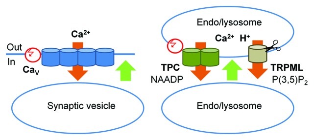
Figure 3. A schematic representation of the role of TPC and TRPML channels in membrane fusion. Left panel: The classic scheme of membrane fusion in presynaptic terminals: Ca2+ influx through the voltage-regulated L-type CaV channels (orange) triggers conformational change in SNARE complexes (not shown) and membrane fusion (green). This influx it triggered by a change in the membrane potential (red). Right panel: Membrane fusion in the endocytic pathway. Ca2+ efflux out of the H+- and Ca2+- rich endo/lysosomes (orange) through TPC and TRPML channels triggers conformational change in SNARE complexes (not shown) and membrane fusion (green). Ca2+ efflux through TPC channels is triggered by NAADP and modulated by membrane potential (red) and Ca2+. TRPML Ca2+ fluxes are activated by PI(3,5)P2 and TRPML1 activity is terminated by cleavage.

An official website of the United States government
Here's how you know
Official websites use .gov
A
.gov website belongs to an official
government organization in the United States.
Secure .gov websites use HTTPS
A lock (
) or https:// means you've safely
connected to the .gov website. Share sensitive
information only on official, secure websites.
