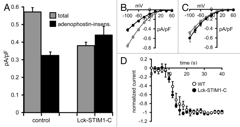
Figure 5. Carbachol-activated currents in HEK cells expressing the Lck-STIM1-C construct. (A) Mean (± SE) carbachol-activated Ca2+ currents measured at -80 mV in HEK cells expressing the m3 muscarinic receptor. Shown are the total agonist-activated currents (gray), and the same currents recorded in cells after depletion of ER Ca2+ stores by intracellular adenophostin A (black) in untransfected control cells (control) and in STIM1 siRNA-treated cells expressing the Lck-STIM1-C construct. (B) Mean (± SE) I/V curves for the total agonist-activated currents (gray), and adenophostin-insensitive currents (black) in control cells and (C) the same for the Lck-STIM1-C expressing cells (see text for details). (D) Mean (± SE) curves comparing the activation kinetics of carbachol-activated currents in m3-HEK cells expressing the Lck-STIM1-C construct compared with those in wild-type m3-HEK cells. Total currents were normalized to their final maximal values to allow direct comparison of their respective rates of activation following addition of carbachol (10 µM) at time zero.

An official website of the United States government
Here's how you know
Official websites use .gov
A
.gov website belongs to an official
government organization in the United States.
Secure .gov websites use HTTPS
A lock (
) or https:// means you've safely
connected to the .gov website. Share sensitive
information only on official, secure websites.
