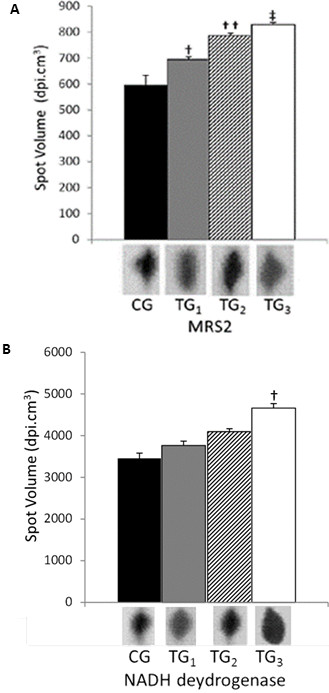
Figure 2.
MRS2 and NADH protein quantity analyses. MRS2 (A) and NADH dehydrogenase (B) expression evaluated by high molecular mass 2-DE technique in rats left ventricle cardiomiocities. CG corresponds to control group; TG1; TG2 and TG3 correspond respectively to 2.5; 5.0 and 7.5 to training groups. Different spots volumes are determinate by using software Bionumerics™. Simbols †, †† and ‡, represent the statistical difference between respectively groups. Statistical analyses were conducted by ANOVA (P < 0.05). All studies were performed in triplicate.

