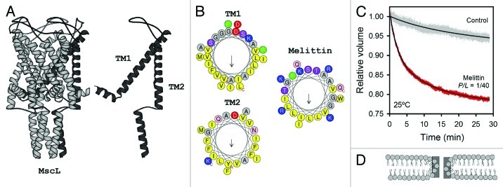
Figure 4. (A) Structure of the mechanosensitive channel MscL (PDB 2OAR, taken from ref. 9). (B) Helical wheel representation for the TM fragments of MscL and a monomer of melittin. Melittin and TM2 were modeled in α-helical wheel representations; since TM1 is larger, it was modeled as a π-helix, which promotes a better lipid exposing face. Hydrophobicity factors (H) obtained are: 0.817 (TM1); 0.959 (TM2) and melittin (0.511). Net charge (z): –1 (TM1); 0 (TM2); + 5 (melittin). Residues with hydrophobic side chains are shown in yellow. Small residues Gly and Ala are represented in gray and positively charged snorkeling residues, in blue. N- and C-ends are shown. Arrows show the hydrophobic moment. (C) Osmotically induced inflow of NH4+ through melittin pores in MLVs of DOPC:DPPC (2:1). MLVs were concentrated at 30 mM in choline chloride. Melittin was added to a P/L ratio of 1/40 in the presence and absence of a hypoosmotic downshock. For the osmotic shock, the sample was diluted 1:70 into an ammonium solution (100 mM). Permeability was determined by the swelling rate for a period of time, measuring the change in turbidity at Abs = 450nm. All flux measurements were performed at 25°C. (D) Schematic drawing of a toroidal pore in a lipid bilayer, taken from ref. 118.

An official website of the United States government
Here's how you know
Official websites use .gov
A
.gov website belongs to an official
government organization in the United States.
Secure .gov websites use HTTPS
A lock (
) or https:// means you've safely
connected to the .gov website. Share sensitive
information only on official, secure websites.
