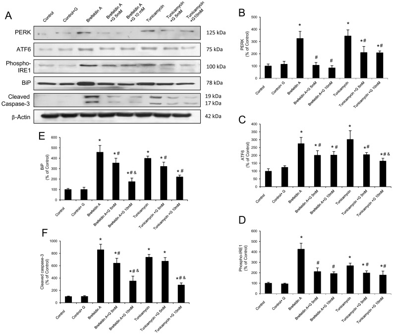
Figure 5. Glutamine reduces the ER stress and apoptosis induced by brefeldin A and tunicamycin in Caco-2 cells.
(A–F) Protein from Caco-2 cells was separated by sodium dodecyl sulfate-polyacrylamide gel electrophoresis, followed by immunoblotting for PERK, ATF6, phosphorylated IRE1 (phospho-IRE1), BiP and cleaved caspase-3. PERK, ATF6 phospho-IRE1, BiP and cleaved caspase-3 were markedly expressed in cells treated with brefeldin A or tunicamycin alone. However, glutamine administration (5 and 10 mM) partially abolished the changes induced by brefeldin A and tunicamycin. Results are representative of four independent experiments. Equal loading of proteins is illustrated by β-actin bands. (A) Representative Western-blot photographs for PERK, ATF6, phospho-IRE1, BiP, cleaved caspase-3 and β-actin. (B) Densitometric quantification of PERK. (C) Densitometric quantification of ATF6. (D) Densitometric quantification of phospho-IRE1. (E) Densitometric quantification of BiP. (F) Densitometric quantification of cleaved caspase-3. Data are expressed as mean ± S.E.M. *P<0.05 compared with control group. #P<0.05 compared with same stress inducer without glutamine group. &P<0.05 compared with same stress inducer +5 mM glutamine group.

