Figure 1.
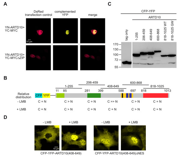
Subcellular localization of ARTD10.A. ARTD10 interacts with the oncoprotein MYC in the nucleus. The indicated fusion proteins were transiently expressed in HeLa cells. The expression of DsRed served as a transfection control. The appearance of yellow fluorescence was monitored by confocal microscopy. B. The scheme summarizes the domain structure of ARTD10 (light green, RNA recognition motif RRM; green, glycine-rich region; yellow, glutamate-rich region; black, nuclear export sequence; blue, ubiquitin-interaction motif; red, catalytic domain). The fusions of full-length ARTD10 and of overlapping fragments of ARTD10 to CFP-YFP are indicated. These fusion proteins were transiently expressed in COS7 cells and their subcellular distribution determined in the presence or absence of Leptomycin B (LMB). The relative distribution between the cytoplasmic and the nuclear compartment is indicated for each fusion protein (for representative micrographs see Figure 2B). C. The fusion proteins indicated in panel B were expressed transiently in COS7 cells and analyzed by Western Blotting using an anti-GFP antibody. D. The subcellular localization of CFP-YFP-ARTD10(408–649) and CFP-YFP-ARTD10(408–649)ΔNES was analyzed in transiently transfected COS7 cells in the presence or absence of LMB using confocal microscopy.

