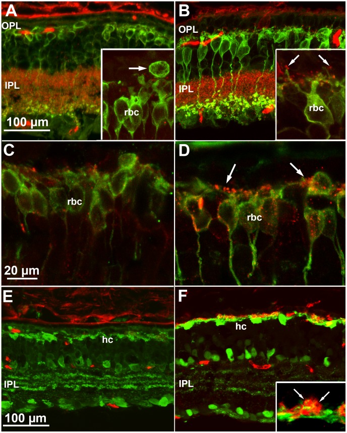
Figure 6. Morphology of inner retinal cells in mice raised in ST and EE.
(A and B) Vertical retinal sections stained with antibodies against PKCα (green), labelling rod bipolar cells (rbc), and with antibodies against bassoon (red) that, in the outer plexiform layer (OPL), label synaptic contacts established by photoreceptors. Age is P60. In ST there is pronounced dendritic retraction of rbc (A) while in EE these are better preserved (B). Also in EE, dendrites are decorated by bassoon-positive puncta (B, arrow in insert) that are virtually absent in ST (A, insert; the arrow points to the displaced cell body of a rbc). (C and D) mGluR6 (red) and PKC staining (green) show that rod bipolar cells, in EE retinas (D) maintain longer dendrites and a complement of mGluR6 (arrows), which are rare in ST counterparts (C). Age is P60. (E and F) Calbindin-D-28 staining of horizontal cells (hc, green). In ST (C), horizontal cell processes in the outer retina are scant and poorly ramified. In contrast, in EE (D), the still branched dendrites of these neurons are surrounded by photoreceptor synaptic endings, labelled by anti-PSD95 antibodies (red), which highlight the round shape of their terminals (inset). Insets are shown at twice the magnification of the main panels.

