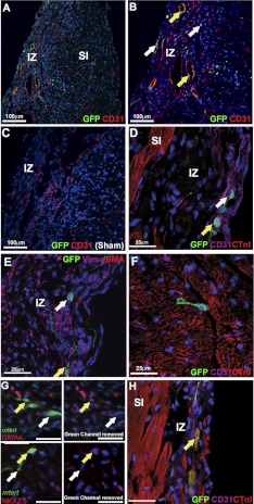
Figure 5.
In the adult heart, all mTert-GFP cell phenotypes described in neonates were found at 14 d after cryoinjury. A) mTert-GFP cells in the injury zone (IZ) and subinjury (SI) zone. B) In the IZ, CD31 expression shows the presence of structures resembling blood vessels, which contain mTert-GFP+ CD31+ cells (yellow arrows). Single mTert-GFP-expressing CD31− cells were also observed in the epicardium and myocardium of the IZ, as well as the SI zone (white arrows). C) Cryoinjured wild-type littermate controls expressed a similar CD31 profile to mTert-GFP animals, but no GFP reactivity was observed. D) mTert-GFP+ CTnI− and mTert-GFP+ CD31− cells were observed in the SI myocardium. E, F) Higher-magnification images of the IZ showing mTert-GFP+ Vim+ (E) and mTert-GFP+ CD31 (F) cells. G) A population of mTert-GFP cells within the IZ express cardiac transcription factors GATA4 and Nkx2.5. H) Cells with mature cardiomyocyte morphologies expressing mTert-GFP and CTnI are found within the IZ (yellow arrows). A–D) Green, mTert-GFP; red, CD31 or CTnI; blue, DAPI (nuclei). E–F) Green, mTert-GFP; red, CTnI, α-SMA, GATA4, or Nkx2.5; magenta, CD31 or vimentin; blue, DAPI (nuclei). White arrows indicate mTert-GFP only; yellow arrows indicate mTert-GFP cells coexpressing investigated markers. Scale bars = 25 μm or as indicated.

