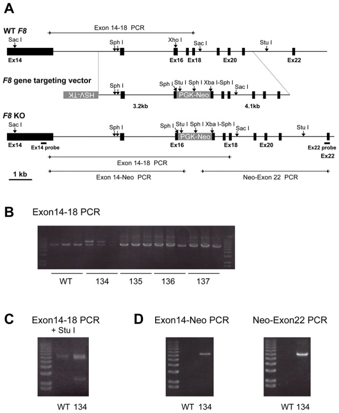
Figure 1. F8 targeting of porcine fetal fibroblasts (PEF).

(A) Schematic diagram of part of porcine F8, the positions of the restriction endonuclease sites, the F8 targeting vector structure, and the targeted F8 (F8 KO) allele are shown. The neomycin-resistance gene (PGK-neo) was inserted in the exon 16 DNA fragment with deletion of a part of exon 16 and was flanked by two F8 DNA fragments (5′ arm: 3.2 kb; 3′ arm: 4.1 kb) in F8 targeting vector. The positions of PCR primers (arrowheads), expected amplified DNA fragments (bars), and restriction endonuclease sites used for the Southern blot analysis are indicated in the schema for F8 KO. (B) F8 exon 14–18 PCR on genomic DNA from non-transfected PEF (WT), PEF colony 134 (134), and three other PEF colonies (135–137) was shown. (C) The F8 exon 14–18 PCR products were treated with Stu I and analyzed by agarose gel electrophoresis. (D) PCR analyses with two sets of primer pairs for exon 14 and the neomycin resistance gene and for the neomycin resistance gene and exon 22 were shown.
