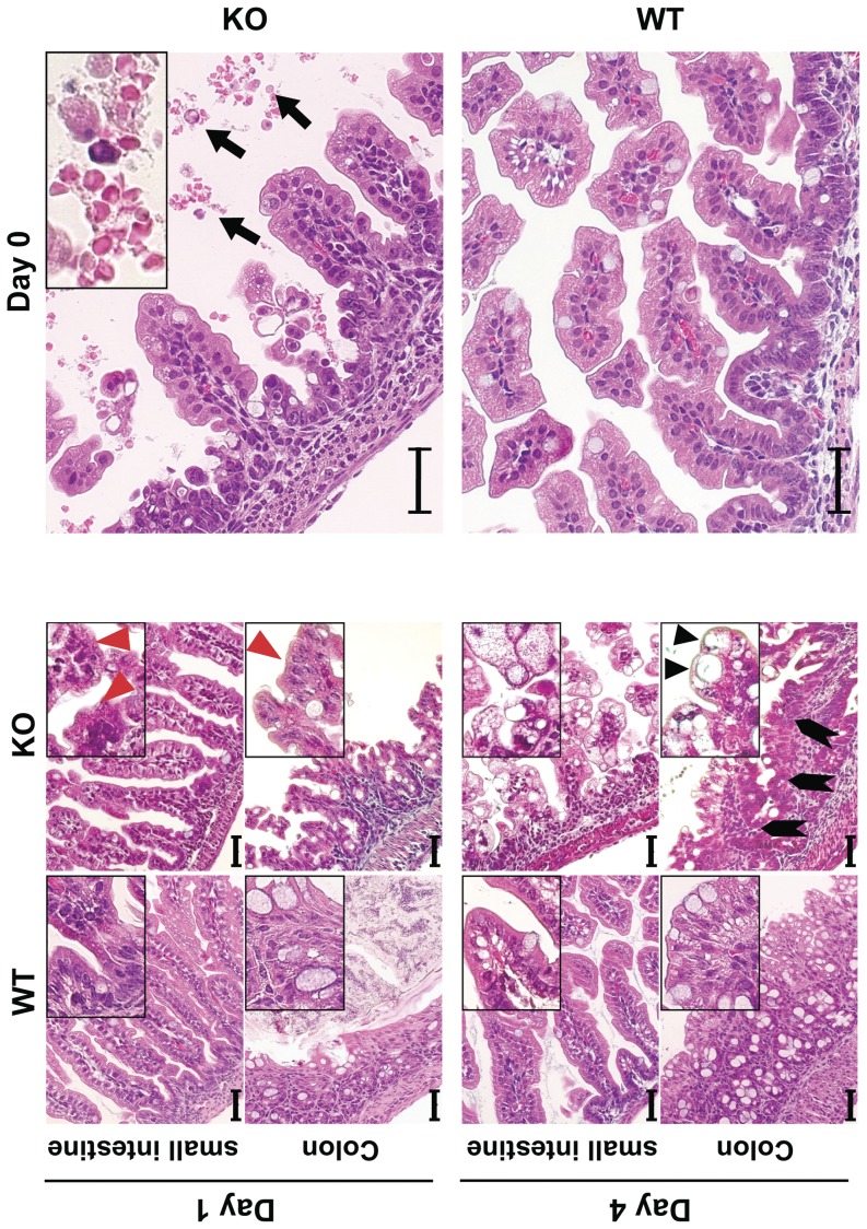
Figure 4. Tufting enteropathy in mTrop1-null mice.
H&E staining of formalin-fixed paraffin-embedded small intestine and colon sections from WT and KO newborn mice, from day 0 to day 4. Insets: magnified areas. Villous atrophy was found throughout the small intestine of KO mice. Severity progressed from day 0 to day 4 (day of death). Red arrowheads: tufts of extruding epithelium, with surface enterocyte disorganization and focal crowding. These abnormalities were focally distributed, and increased over time, with highest tuft density at the time of death. Lymphocytes and plasma cells in the lamina propria were infrequent. KO colon crypts showed pseudo-cysts formation (black arrowheads) and abnormal regeneration with branching (block arrows). Hemorrhagic enteritis was apparent in the small intestine of KO mice from day 0 (top, right); black arrows: red blood cells in the intestinal lumen. Scale bars: 40 µm.

