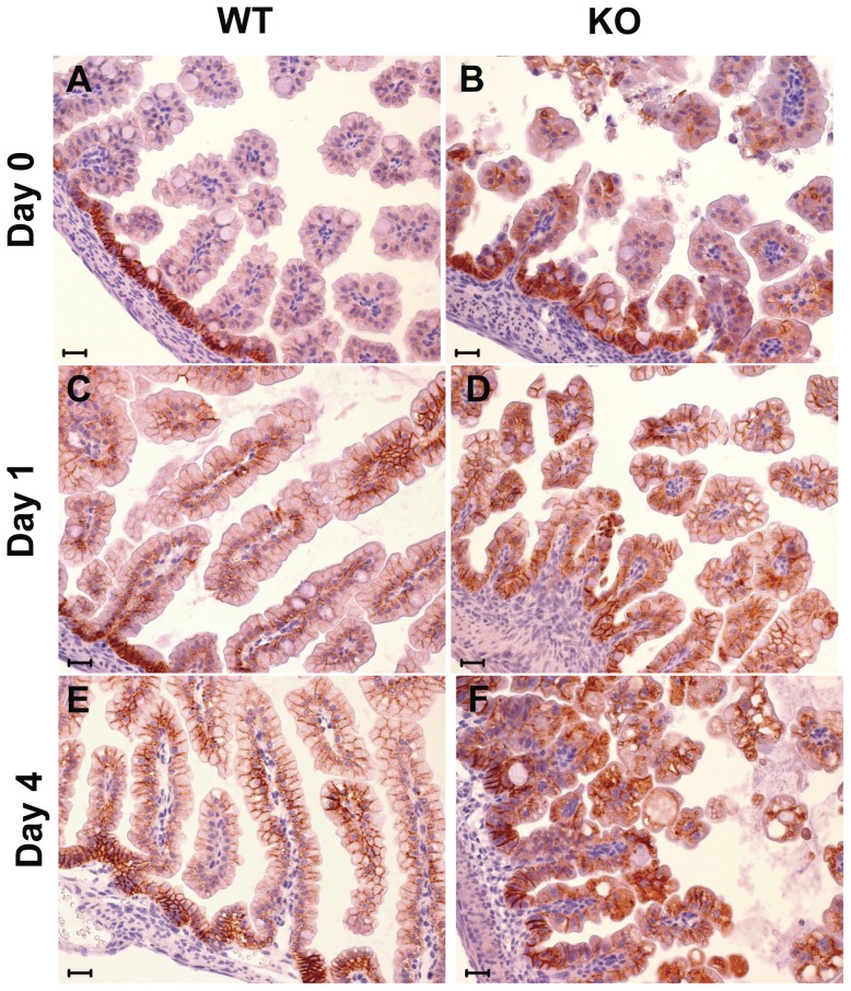
Figure 6. E-cadherin expression in the intestine.
E-cadherin immunostaining pattern in small bowel of WT (wt) and KO mice at birth (day 0), and at day 1 and day 4 after birth. E-cadherin shows a typical membrane immunoreactivity in WT mice (A, C, E), whereas in KO mice (B, D, F) it is localized increasingly in the cytoplasm, with a prevalent cytoplasmic accumulation and membrane-disrupted pattern at day 4 after birth. (Scale bar: 20 mm).

