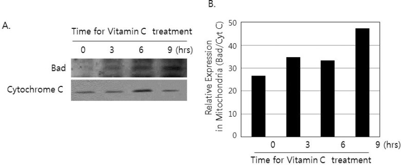
Figure 4.
The translocation of Bad from 14-3-3β to mitochondria by the treatment of vitamin C. (A) Cells (2×10⁶) was incubated for 3, 6 and 9 hrs in the presence or absence of 2 mM of vitamin C. Mitochondrial fraction was prepared as described in materials and methods. Then western blotting was done by using antibodies against Bad and cytochrome C. (B) The density of each band was measured by densitometry, and the values were expressed as the ratio Bad/Cytochrome C.

