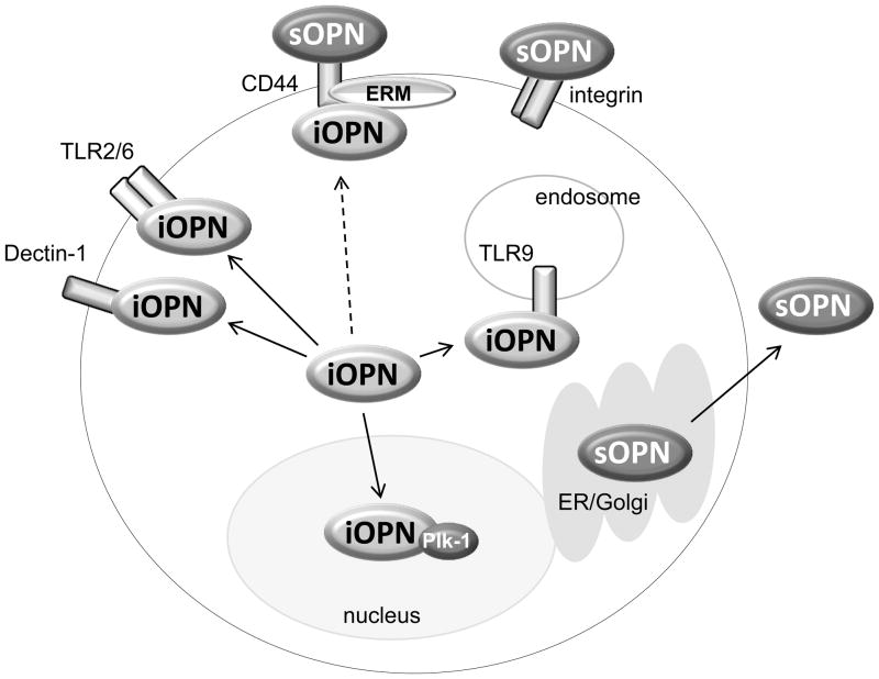
Fig. 1. Interaction of OPN and other molecules.
In unstimulated cells, iOPN appears to be distributed in cytoplasm of macrophages and dendritic cells. Once cells are stimulated with pattern-recognition receptor ligands, iOPN is recruited to the receptor that detects a ligand. Zymosan and fungal stimuli recruit iOPN to molecular complexes downstream of TLR2 (fungal-recognizing TLR2 makes heterodimer with TLR6) and dectin-1 (Inoue et al. Submitted). CpG DNA stimulation make iOPN to translocate to TLR9 (Shinohara et al., 2006). iOPN is also known to co-localize with CD44 and ezrin/radixin/moesin (ERM) protein ezrin (Zohar et al. 2000), although it is not known whether the colocalization is activation-specific (broken arrow). iOPN translocates into nuclei particularly during the G2/M period of mitosis, and interacts with Plk-1 (Junaid et al. 2007). sOPN is secreted through ER and Golgi. Receptors of sOPN are CD44 and various integrins.

