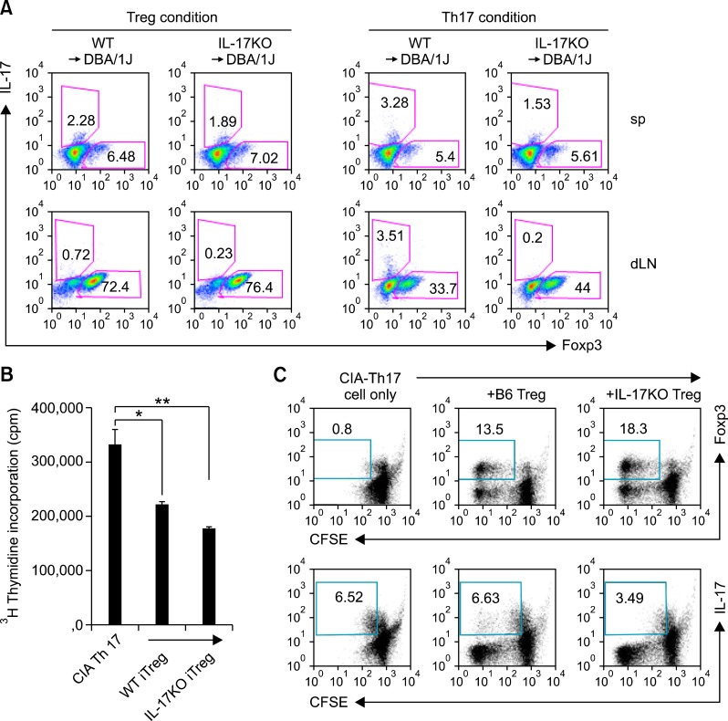
Figure 4.
Inhibition of effector T cell proliferation/response by interleukin (IL)-17 knockout (KO) bone marrow transplant (BMT)-induced Tregs. (A) CD4+ T cells were isolated from both chimeric mice, C57BL/6 (B6 → DBA) or IL-17-/- bone marrow (IL-17-/- → DBA), and cultured under activating conditions with Treg or Th17 polarizing conditions for 3 days, respectively. [Treg condition (Left): anti-CD3 and anti CD28 in the presence of recombinant hTGF-β, Th17 condition (Right): anti-CD28 in the presence of recombinant IL-6, TGF-β, anti-IL-4, and anti-IFN-γ]. Representative FACS plot depicting Foxp3 or IL-17 expression by CD4+ T cells. (B) CD4+ CD25+ T cells sorted from the above Treg condition cell cultures (a, left) were assessed for their suppressive capacity. CD4+ CD25+ T cells (5 × 10⁴ cells) were cultured with irradiated (5,000 rad) APCs (5 × 10⁴ cells) and Th17-conditioned culture T cells (5 × 10⁴ cells) isolated from CIA mice in the presence of CII for 72 h. The proliferation of effectors (Th17-conditioned T cells) was measured by incorporation of 3H-thymidine. Values are presented as means ± SEM. *P < 0.05, **P < 0.01. Data shown are representative of three independent experiments. (C) The regulatory effect of Treg in each group of mice. Treg were generated in vitro, sorted to > 99% purity, and cultured with effector cells. Treg cells from each group were cultured with CFSE-labeled CD4+ effector T cells from CIA mice. After 3 days, proliferation of effector cells co-cultured with Treg from B6 or IL-17KO was measured by CFSE dilution. Percentages of dividing effector cells (determined by CFSE incorporation) are shown. Data shown are representative of three independent experiments.

