Figure 5.
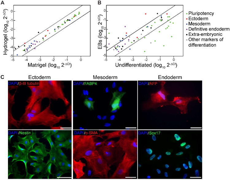
Long-term culture of hES cells on 10 kPa hydrogels. A. Gene expression analysis of hES cells (H9) cultured for 60 days on hydrogels functionalized with CGKKQRFRHRNRKG using quantitative PCR (qPCR). The level of gene expression is compared to cells cultured on Matrigel-coated plates. B. Gene expression analysis of embryoid bodies generated from the long-term (60 days) cultured hES cells. The level of gene expression is relative to undifferentiated cells cultured on the peptide-bearing hydrogels. For both qPCR scatter plots, black lines represent a four-fold increase or decrease between control and experimental conditions. C. Microscopy images of embryoid bodies developed from long-term (38 days) cultured cells that were immunostained for markers of all three embryonic germ layers; ectoderm (nestin and β-III tubulin), mesoderm (FABP4 and α-SMA), and endoderm (AFP and Sox17). Scale bars: 100 μm for nestin, 50 μm for all others.

