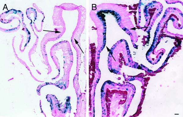
Figure 1.

No-air and air-assisted infected epithelia
A) Cryosections of no-air and B) air-assisted epithelium infected with Ad-lacZ and counterstained with eosin. Air-assisted epithelia (n=9) show significantly increased rates of infection throughout the epithelium compared to no-air (n=6). Expression can also be detected in the dorsal recess (compare area between black arrows in (A) and (B)). Scale bar = 100 μm.
