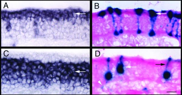
Figure 2.

Infection of sustentacular cells and OSNs by Ad-lacZ
A) In situ hybridization with cytokeratin 18, which labels sustentacular cells primarily at the apical surface (arrow). B) LacZ activity detected in the sustentacular layer in Ad-lacZ infected mice. Infected cells have processes that extend to the basal surface (arrow). C) In situ hybridization with OMP, which labels mature OSNs (arrow). D) LacZ activity detected in cells located within the olfactory nerve layer (white arrow). Dendritic processes of OSNs extending to the apical surface (black arrow) as well as axonal projections (white arrowhead) can be detected. Scale bar = 20 μm.
