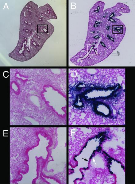
Figure 4.
Infection of proximal and distal respiratory tract by Ad-lacZ
A) Cryosection of lungs from an animal infected with Ad-lacZ using the no-air instillation method. B) Cryosection of an animal infected using the air-assisted method. C) Area in (A) outlined by black box. D) Area in (B) outlined by black box. E) Area in (A) outlined by white box. F) Area in (B) outlined by white box. Variability in lacZ staining is observed in the most distal bronchioles (F), with stained (white arrow) and unstained (black arrow) cells. LacZ staining could be seen in these sections from no-air instilled animals (C,E), but the expression was extremely weak. Scale bar = 640 μm (A,B) and 100 μm (C-F).

