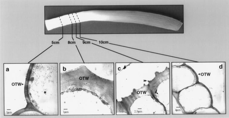
Figure 5.
Transmission electron micrographs of epidermal cells at various positions along the length of the leaf. a, Abaxial epidermal cells at 5 cm above the base of the leaf showing the thin OTW and smooth cuticular surface. The cuticle is a thin line on the surface of the OTW. b, Abaxial (outer) epidermal cells at 8 cm showing the thickened OTW and a single central ridge as the beginning of cuticular surface irregularities. The single ridge was observed on the cuticular surface of all abaxial epidermal cells examined in this region. c, Abaxial epidermal cells at 9 cm showing the increasing irregular cuticular surface and the pair of developing ridges over the radial cell walls (double arrowhead). d, Adaxial (inner) epidermal cells at 10 cm showing the thin cuticle and the OTW, which was thin and smooth at the cuticular surface. The epidermal cells on the abaxial surface at 9 cm had already undergone more extensive morphological changes (shown in c) and had a heavy deposit of wax crystals. The adaxial surface did not have wax crystals in this region, although dense wax crystals were evident in segments from higher up on the leaf. Scale bars are indicated.

