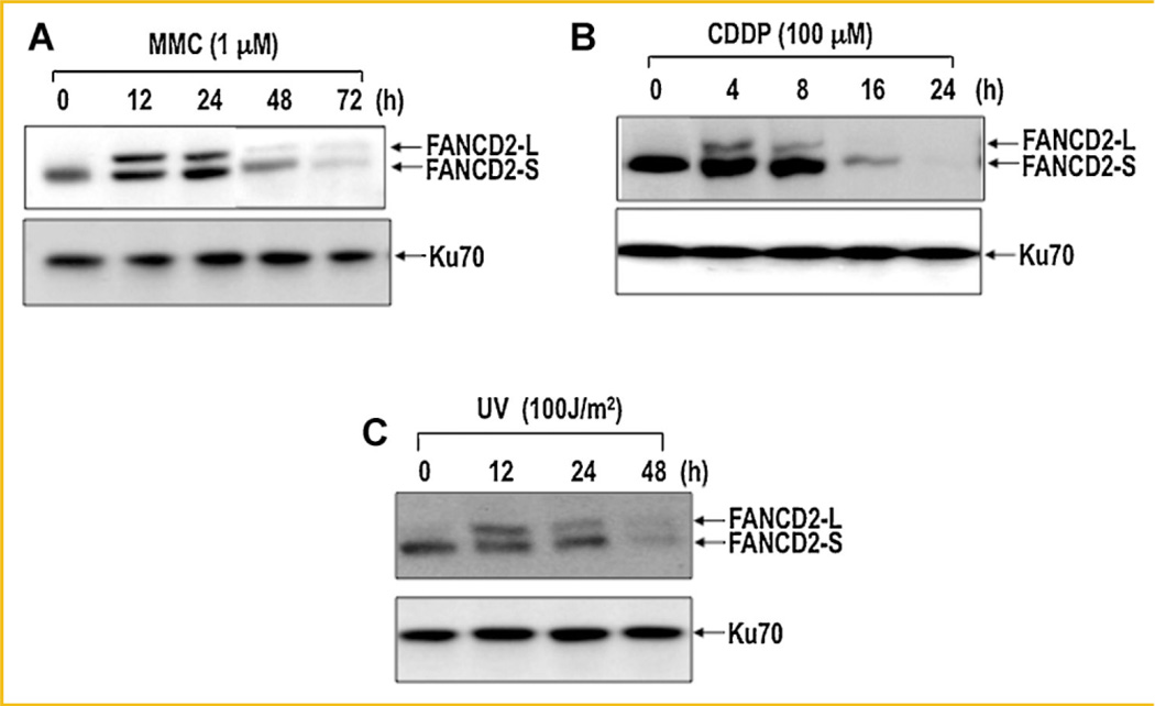
Fig. 1.
Disappearance of FANCD2 occurs following treatment of cells with DNA damaging agents. HeLa monolayer cells with 50% confluence were treated with 1 µM mitomycin C (MMC, panel A), 100 µM cisplatin (CDDP, panel B), or 100 J/m2 germicidal UV light (panel C). Cells were harvested at various time points and analyzed for FANCD2 by Western blot. FANCD2-L and FANCD2-S represent monoubiquitinated and non-ubiquitinated forms of FANCD2, respectively. Ku70 was used as an internal loading control for disappearance of FANCD2.

