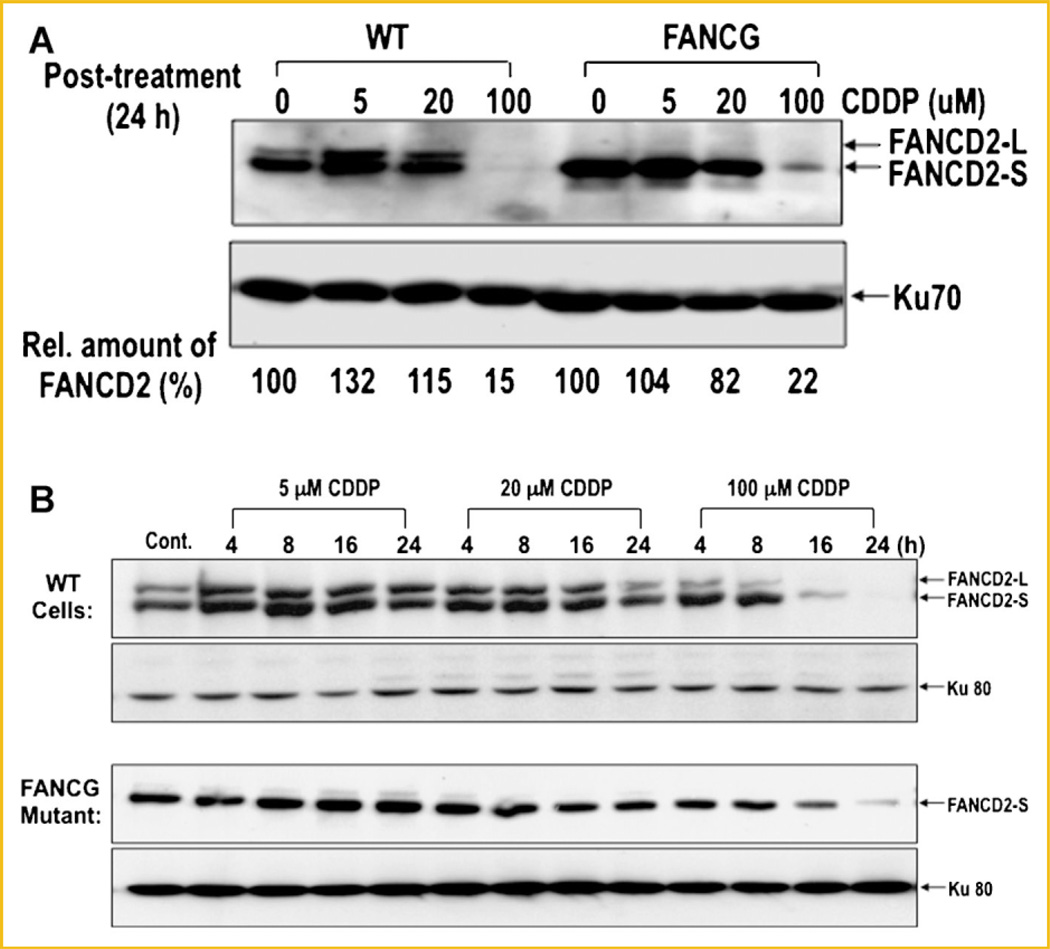
Fig. 6.
Disappearance of FANCD2 in WT cells and FANCG mutants following CDDP treatment. A: WT fibroblast and FANCG mutant cells were analyzed for disappearance of FANCD2 protein following CDDP treatment for 24 h. Cell extracts were subjected to 8% SDS–PAGE followed by Western blot using an anti-FANCD2 antibody. Ku70 was used as a loading control. Relative amount of FANCD2 protein (%) was quantified from theWestern blot (top panel) using the NIH image system. B: Kinetic analysis of disappearance of FANCD2 in WT cells and the FANCG mutant following CDDP treatment. Cells were treated with indicated amount of CDDP, harvested at various times, and analyzed for relative amounts of FancD2-L and FancD2-S by Western blot.

