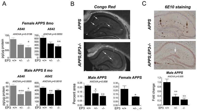
Figure 4. Deletion of EP3 receptor in APPS mice reduces levels of Aβ40 and Aβ42 peptides.
(A) Levels of total Aβ40 and Aβ42 were measured in female and male 8 mo APPS;EP3 cohorts. Total Aβ40 and Aβ42 levels were significantly lower in APPS mice lacking one or both alleles of EP3 in both genders (females: n=10–11 per group, ANOVA p<0.05 for Aβ40 and p<0.01 for Aβ42 levels; males: n=9–15 per group; ANOVA p<0.001 for Aβ40 and p<0.01 for Aβ42 levels). (B) Staining of hippocampus with Congo Red (left panels, diagonal arrows) and quantification of Congo red positive percent area demonstrates a lower level of Aβ peptide β-pleated sheet in 8 mo APPS;EP3+/− and APPS;EP3−/− male mice (n=3–4 mice per genotype), and in 8 mo combined APPS;EP3+/− with APPS;EP3−/− female mice (n=4 APPS;EP3+/− mice combined with n=2 APPS;EP3−/− mice; n=8 APPS mice; *p<0.05 and ** p<0.01). (C) Quantification of 6E10 immunostaining in hippocampus demonstrates significant decreases in amyloid plaque deposition (vertical arrows) in 8 mo male APPS mice lacking one or both alleles of EP3 (n=3–6 per group for males ANOVA p<0.01, with post-hoc p<0.05 and **p<0.01 relative to APPS genotype).

