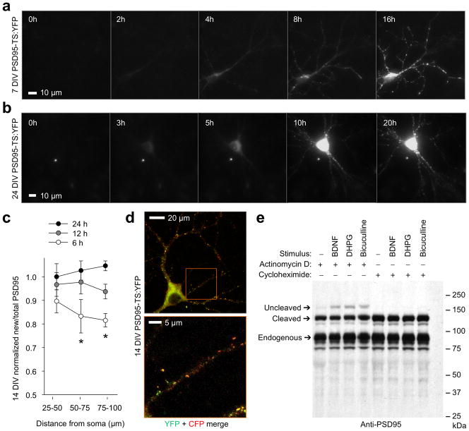
Figure 2.
Tracking of basal and activity-induced PSD95 production with TimeSTAMP. (a) New PSD95 accumulates in dendritic puncta during arborization in a 7-DIV neuron. New puncta appear on distal branches simultaneously with their extension. Times in 1 μM BILN-2061 are shown. (b) At 21 DIV, new PSD95 accumulates in puncta throughout the dendritic tree. (c) Distally located synapses incorporate less new PSD95 than proximal ones in 14-DIV neurons. PSD95-TS:YFP fluorescence in segments of the primary dendrite was divided by cotransfected PSD95-CFP fluorescence for each time, then normalized to the initial value in the 25-μm segment. Mean differences between times were significant for 50–75 μm and 75–100 μm segments by ANOVA (p = 0.018 and 0.020 respectively, n = 7 segments each). Differences between 24 h and 6 h were significant by post-hoc Tukey’s tests (p = 0.017 and 0.016 for segments at 50–75 μm and 75–100 μm, respectively). Error bars represent SEM. (d) Upper image, a representative neuron from (c) showing that PSD95 synthesized for 20 h in 1 μM BILN-2061 under basal conditions exists in a gradient from the cell body. PSD95-TS:YFP protein is in green while PSD95-CFP, a marker of total PSD95, is in red. Lower image, enlarged view of boxed region in upper image. (e) TimeSTAMPa revealed induction of PSD95 synthesis by stimuli. 14-DIV neurons expressing PSD95-GFP-TimeSTAMPa were treated for 3 h with 10 μM BILN-2061 and stimuli along with actinomycin D to block transcriptional upregulation or cycloheximide to block protein synthesis. Anti-PSD95 immunoblotting reveals proteins synthesized during BILN-2061 incubation migrating at the uncleaved size. 100 ng/mL BDNF, 50 μM DHPG, and 20 μM bicuculline each induce PSD95 synthesis above baseline independent of transcription. Endogenous PSD95 serves as a loading control.

