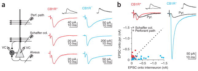
Figure 3.
Distinct excitation of CB1R-positive and -negative basket cells. (a) Left, schematic of recording configuration. Monosynaptic EPSCs recorded in a CB1R-positive (middle) and -negative (right) basket cell by stimulating three excitatory pathways. (b) Top, EPSC recorded simultaneously in connected basket-to–pyramidal cell pairs in response to Schaffer collaterals stimulation. Same cells as in a. In a and b, EPSCs were recorded in the presence of gabazine (2.5 μM) or at the IPSC reversal potential (−85 mV). Bottom, scatter plot of the amplitude of Schaffer collateral and perforant path EPSCs recorded in CB1R-positive (Schaffer collaterals, n = 16; Perforant path, n = 7) and -negative (Schaffer collaterals, n = 16; Perforant path, n = 5) versus their paired pyramidal cells. Dotted line, unity.

