Figure 4.
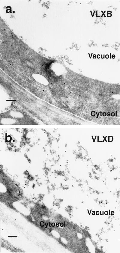
Transmission electron microscopy immunocytolocalization of VLXB and VLXD protein in mature soybean leaves after 4 weeks of continuous pod removal. a, Portion of a leaf cross-section from a plant with pods removed daily for 4 weeks and incubated with anti-VLXB antibody. Tissue sections are identical to those described for Figure 3. Sections were mounted onto nickel grids, incubated with peptide-specific antibody and protein A-gold conjugate, and poststained. Immunogold particles are localized primarily in the cytosol in this cell from a sink-regulated plant. Scale bar = 200 nm. b, Portion of a leaf cross-section from a plant with pods removed daily for 4 weeks and incubated with anti-VLXD antiserum. Tissue sections are identical to those described for Figure 5 and were prepared as in a. Immunogold particles are dense in the vacuole and localize strongly to electron-dense flocculent material in this cell from a sink-regulated plant. Minimal localization is apparent elsewhere. Scale bar = 200 nm.

