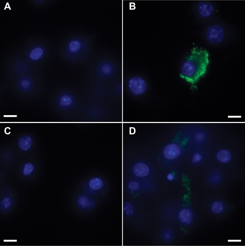
FIG 3.

Riboswitch-mediated control of GFP (GFP-neg) in F. novicida during macrophage infection. RAW264.7 murine macrophages were infected at an MOI of 50:1 with F. novicida harboring gfp lacking a promoter (GFP-neg; panel A) or constitutively expressing gfp (GFP-pos; panel B) or the E-Rs-GFP riboswitch construct (panels C and D). At 30 min postinfection, medium without theophylline (panels A to C) or with 1 mM theophylline (panel D) was added and the macrophages were incubated at 37°C for 24 h and fixed. GFP (green) and DAPI-stained macrophage nuclei (blue) are shown. The magnification is ×100, and the scale bar represents 10 µm. The data are representative of three independent experiments in which at least 10 fields of view were analyzed for each condition.
