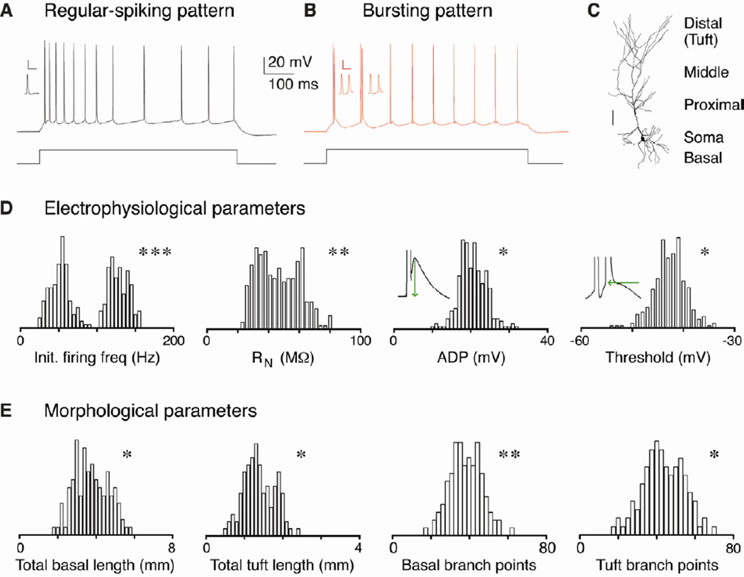
Figure 1. Distribution of electrophysiological and morphological properties.
A-B. Pyramidal neurons in CA1 and the subiculum respond to step current injection in vitro by firing action potentials in one of two distinct patterns: regular spiking or bursting. The former pattern consists of trains of individual action potentials, while the latter pattern begins with one or more bursts of high frequency (>100 Hz) spikes. Inset: enlarged bursts (scale bar = 50 mV and 20 ms). C. Filled pyramidal neurons with different dendritic compartments indicated (scale bar = 100 µm). D-E. All-point histograms of the distributions of several electrophysiological (n = 268 cells) and morphological properties (n = 110 cells). Illustrations of how ADP and second spike threshold were measured are inset. P values from the D’Agostino & Pearson omnibus normality test demonstrate that the properties presented are not unimodally distributed, suggesting that there are discrete groups of pyramidal cells within this population. Asterisks indicate significant differences between the groups (*p < 0.05, ** p < 0.01, *** p < 0.001).

