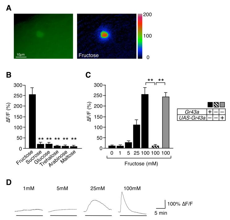
Figure 3. GR43a functions as a fructose sensor in the brain.
(A) Brain neuron expressing G-CaMP3.0 under control of Gr43aGAL4: ΔF pseudocolor fluorescence image was taken 24 seconds after application of 100mM fructose (right).
(B)Gr43aGAL4 neurons specifically respond to fructose. Max ΔF/F within 15 minutes of application is shown. All sugars are 100 mM. Flies contained two genomic copies of Gr43a. **p < 0.0001; ANOVA. Error bars represent standard error. 6≤n≤7.
(C) Response of Gr43aGAL4 neurons to fructose is dose- and Gr43a dependent. **p < 0.0001; ANOVA. Error bars represent standard error. 8≤n≤9.
(D) Time-course of G-CaMP3.0 fluorescence changes in Gr43aGAL4 neurons stimulated with different concentrations of fructose.

