
FIG 4 .
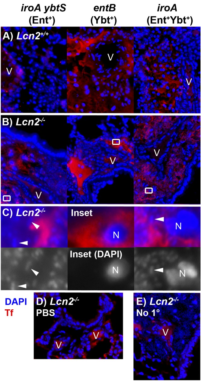
The perivascular spaces contain serum exudate as indicated by transferrin. Merged immunofluorescence images of formalin-fixed paraffin-embedded histological sections of lungs from Lcn2+/+ (A) or Lcn2−/− (B) mice 3 days after retropharyngeal inoculation of 1 × 10⁴ CFU/ml of K. pneumoniae siderophore mutants, stained using antitransferrin rabbit polyclonal IgG (Tf) (red) and the nucleic acid stain DAPI (blue), are shown. Magnified merged and DAPI-only images (C) of insets from Lcn2−/− mice in panel B demonstrate nuclei (N) and collections of bacteria (representative bacilli are indicated by arrowheads) in the perivascular space. Merged images of Lcn2−/− mice mock infected with PBS (D) or infected with iroA mutant K. pneumoniae but lacking antitransferrin antibody (E) are shown as controls. Magnification, ×400, except panel C (×4,000); V, vessels.