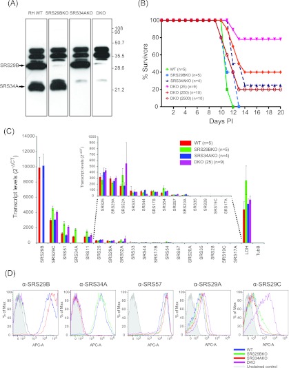
FIG 6.
Targeted disruption of SRS29B and SRS34A alters SRS29C expression and acute virulence in mice. (A) Anti-CRD Western blot profile of Srs29B−, Srs34A−, and DKO parasite strains to identify relative expression levels of SRS antigens expressed on knockout and WT tachyzoites. A total of 107 tachyzoites were solubilized in 1% NP-40, incubated in the presence of GPI-PLC purified from T. brucei for 1 h at 37°C, separated by 12% SDS-PAGE, and transferred to nitrocellulose. SRS proteins were detected by Western blotting with a rabbit anti-CRD polyclonal antibody, followed by a horseradish peroxidase-conjugated anti-rabbit secondary antibody, and enhanced chemiluminescence. (B) Death kinetics of seropositive CD-1 mice infected with engineered strains of T. gondii. The numbers of seropositive mice infected with the WT, Srs29B−, and Srs34A− strains were five, five, and four, respectively. The total infectious dose was 25 parasites. The numbers of seropositive mice infected with the DKO strain at infectious doses of 25, 250, and 2,500 parasites were 9, 10, and 10, respectively. Mortality data are a composite of three independent experiments with independently derived knockout parasite clones. (C) Gene expression was measured by TaqMan qRT-PCR. The experiment shown is for one representative data set from two independent mRNA-cDNA extractions. Data are clustered from most abundant (SRS29B) to least abundant (SRS17A) transcripts. Transcript levels are represented as 2−∆∆CT to show absolute levels of transcripts relative to every SRS gene examined and were normalized against 18S rRNA genes transcripts. (D) Relative levels of surface-expressed SRS proteins on WT, Srs29B−, Srs34A−, and DKO parasites were determined by flow cytometry by using DG52, 5A6, 4F12, SUS1, and CL15/4 to detect SRS29B, SRS34A, SRS57, SRS29A, and SRS29B, respectively.

