
Fig. 2. Whole mount miRNA in situ hybridization on embryos.
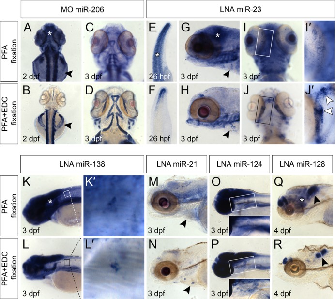
Top rows of top and bottom panel: miRNA in situ hybridization on embryos fixed in PFA only. Bottom rows of top and bottom panel: miRNA expression detected in embryos fixed in PFA+EDC. All embryos where processed simultaneously and stained for approximately 24 h. All arrowheads indicate miRNA expression detectable with high resolution in PFA+EDC fixed samples versus fixation in PFA only. Asterisks indicate non-specific background signals in PFA fixed embryos (A,E,G,K,Q). (A–D) MiR-206 expression in skeletal muscle cells was detected by a CF-labeled MO probe at 2 dpf (A,B) and 3 dpf (C,D). Arrowheads (A,B) point to skeletal muscle cells located on the yolk that were detectable in PFA+EDC fixed embryos (B) and not in PFA fixed embryos (A). (E–J′) MiR-23 is expressed in the tail tip at 26 hpf (E–F), in the cardiac cushions at 3 dpf (G,H, arrowheads) and in developing bone structures of the jaw at 3 dpf (I–J′). (I′–J′) Magnification of boxed areas in I and J show enhanced miR-23 expression in future joints (arrowheads in J′), which was not detectable in PFA fixed embryos (I′). (K–L′) miR-138 is expressed in the developing brain (K,L) and motor neurons (K′,L′). (M,N) MiR-21 is expressed in cardiac cushions (arrowheads) at 3 dpf in PFA+EDC fixed embryo (N), which was not detectable in PFA fixed embryos (M). (O,P) Expression of miR-124 at 3 dpf in the brain and motor neurons located ventrally of the developing brain (boxed area) is hardly detectable in PFA fixed embryos (O) while clearly visible in PFA+EDC fixed embryos (P). (Q,R) Highly specific expression of miR-128 in two separated regions of the developing brain is seen in PFA+EDC fixed embryos at 4 dpf (arrowhead in R) compared to more diffuse staining in PFA fixed embryos (arrowhead in Q).