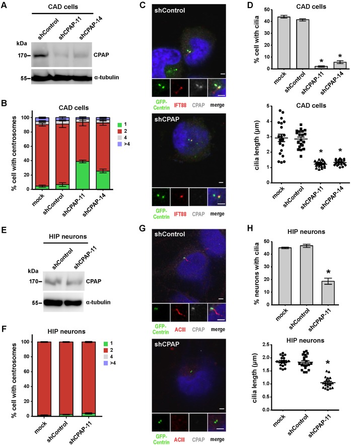
Fig. 2. Depletion of CPAP suppresses cilia formation.
CAD cells (A-D) and hippocampal neurons (E-H) were transfected with shControl or shCPAP (-11 and -14) constructs and then serum starved for 72 hours (CAD cells) or 4 DIV (hippocampal neurons). Western blot analysis of shCPAP knockdown in CAD cells (A) and hippocampal neurons (E); α-tubulin was used as a loading control. Quantitative analysis of centrosome numbers in CAD cells (B) and hippocampal neurons (F) treated with shCPAP. Immunofluorescence images of shControl- and shCPAP-11-treated CAD cells (C) and hippocampal neurons (G) labeled with DAPI and the indicated antibodies. Quantitative analysis of ciliated cell numbers and cilia length in CAD cells (D) and hippocampal neurons (H). Bar values are means ± s.d. of three experiments (n = 100 counted for ciliated cells; n = 20 counted for cilia length). *p<0.05 vs. the respective control (Student's t-test). Due to the expression of exogenous shRNA is gradually reduced in hippocampal neurons after long-term culture (> 5 DIV), we selected 4 DIV as the time point for analysis. Bar in C,G: 2 µm.

