
Fig. 3. The Wolbachia are absent from the gonadal primordium in B. malayi male and female L3 larvae.
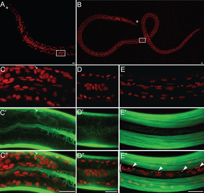
B. malayi L3 larvae stained with PI -red- and phalloidin -green-. (A) anterior part of a female L3 larva and (B) male L3 larva, all obtained from mosquitoes 14 days after their mf-infected blood meal. The gonadal primordium (GP, white box) is anterior in the female and in the mid-body in the male. The anterior is marked by an asterisk. (C–C″) female GP (arrow pointing toward the anchor cell) located at the junction between the developing oesophagus and intestine, and apposed to the hypodermis. (D–D″) male GP. (E–E″) surface detail in the posterior half of a L3 larva, where the Wolbachia (arrowheads) localize in a lateral hypodermal chord (bracket). All the images are merges of confocal sections, the anterior of the worms being oriented to the left. Scale bar = 10 µm.