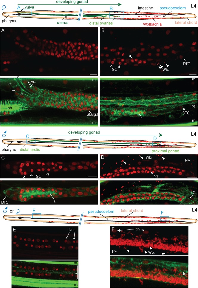
Fig. 4. The Wolbachia start invading the female ovary distal tips from the adjacent posterior lateral chords in L4 larvae, but are absent from the L4 male gonad.
B. malayi L4 larvae (8 to 10 dpi) stained with propidium iodide -red- and phalloidin -green-. The localization of the images taken is indicated on the schematic drawings. (A,B) anterior -proximal- part, and posterior -distal- part of the female gonad respectively. (C,D) anterior -distal- part and posterior -proximal- part of the male gonad respectively. (E) Top down view of a typical anterior lateral chord. (F) Longitudinal view of a typical posterior lateral chord. DTC, distal tip cell; GC, germ cells; i., intestine; sg., somatic gonad; lc., lateral chord; lcn, lateral chord nuclei; LC, linker cell; m., muscle; ps., pseudocoelom; r., rachis; ut., uterus; v., vulva; vc, vulval cells; Wb, Wolbachia; All images are oriented with the L4 larvae anterior part to the left. Scale bar = 20 µm.

